


Delayed detection
Potentially malignant disorders can be subtle, especially when symptoms are not severe enough to trigger specialist referral.
Patent granted
AI point-of-care
NHM pilot tested
Non-invasive screening
AI Oral Cancer Screening
Camera & Optics
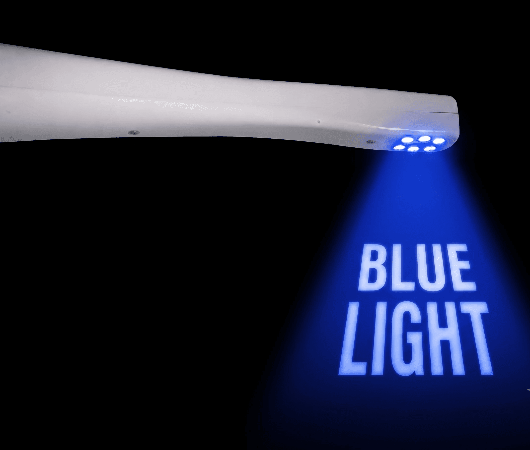
Dr.Oroscope pairs a 5MP HD sensor with a narrow intraoral head designed to access the retromolar region, posterior buccal mucosa, and restricted-opening cases like OSMF and squamous cell carcinoma. Blue illumination in the 400–460nm range — alongside green and amber comparison — reveals tissue changes, lesion margins, and areas that are difficult to access with standard intraoral cameras. Adjustable intensity manages reflection from saliva and adjacent enamel.


Controls & Workflow
Three on-wand buttons handle every clinical input — toggle through white, blue, purple, and green-amber illumination, capture stills, and power the device single-handed. Type-C streams images directly to the connected Android tablet, where point-of-care software supports diagnostic review, registry storage, and telemedicine escalation when a case needs specialist eyes.
IMAGING MODES
Dr. Oroscope captures the same lesion under three distinct illumination modes — giving the AI engine the spectral inputs needed to separate inflammation from dysplasia.
PRECISION VISUALIZATION
For clear surface-level screening.

FLUORESCENCE IMAGING
Highlights mucosal abnormalities under autofluorescence.
SPECTRAL CONTRAST
Highlights hemoglobin-related contrast.
Population-level oral screening is difficult when access to specialists is limited, visual interpretation varies by examiner, and high-risk lesions are often noticed late.
Potentially malignant disorders can be subtle, especially when symptoms are not severe enough to trigger specialist referral.
Traditional visual checks depend heavily on training, lighting, experience, and the ability to evaluate hidden oral areas.
Screening camps, colleges, hospitals, and government programs need tools that are portable, repeatable, and easy to train.
A portable intraoral screening system built for clinics, institutions, camps, and public-health programs.
Visible-Light Imaging
Dr. Oroscope uses blue light in the 400-460nm range with green and amber light comparison to help visualize tissue changes, lesion margins, and hidden areas of the oral cavity. Adjustable intensity and a compact intraoral head make screening easier in posterior regions and limited mouth opening cases.


AI + Telemedicine
Captured images can be processed by software for point-of-care diagnostic support, stored on connected Android/tablet/computer workflows, and escalated through telemedicine support when a case needs specialist review.
A clear documentation trail supports institution-level conversations, from product IP and Startup India recognition to CDSCO/DCGI correspondence and ethical approvals.
Patent Office, Government of India. Portable intraoral cancer screening device, Design No. 394910-001.
NOC correspondence from the Medical Device Division supports the regulatory documentation story.
RAAMAH BIO CARE Pvt. Ltd. recognized in Healthcare & Lifesciences / Medical Devices Biomedical.
Documentation includes institutional ethics and study approvals for validation work.
Supporting research connects Dr. Oroscope with SNP genotyping, pharmacogenomics, and personalized oral medicine for future clinical decision support.
Research material connects oral lesion evaluation with genetic variation and disease-risk understanding.
Pharmacogenomics content supports a future-facing story around therapy selection and reduced trial-and-error care.
AI screening, patient history, images, and specialist support create a stronger referral and follow-up pathway.
Evidence & Deployment
A concise evidence layer for buyers and program teams: publication data, MNJ Institute of Oncology and SVSIDS Mahabubnagar validation, private clinic and college use, NHM pilot deployment, outreach teams, NGOs, and measurable lesion discovery.
0
0
Published study comparing Dr. Oroscope with VELscope for oral cancer screening support.
0+ sites
0+ sites
Multi-center evaluation across key Andhra Pradesh sites, with deployment support from clinical institutions, outreach teams, and NGOs.
0+
0+
People screened across Andhra Pradesh and Telangana through clinical and outreach deployments.
~0
~0
Oral lesions identified during screening, supporting earlier documentation, review, and referral.
The product is designed to work across private care and public-health deployment, with workflows that support capture, reporting, review, and referral.
Chairside screening, patient education, documentation, and referral support.
Portable workflow for high-risk communities and outreach programs.
Teaching, documentation, research, and multi-center validation use cases.
Oral medicine, oncology referral, and follow-up documentation pathways.
Population screening support with reporting and telemedicine potential.
Store, transfer, review, and escalate doubtful cases to specialists.

Capture intraoral images with the compact handheld device, even in hidden or posterior areas.
Use blue light and green/amber comparison modes to visualize tissue changes beyond normal light.
Let the software process images, filters, history, and lesion characteristics for AI-assisted support.
Generate point-of-care reports and escalate doubtful cases through telemedicine support.
Discuss a clinical demo, institutional evaluation, or screening-program rollout with a patent-granted AI oral screening platform.